Eye Conditions We Diagnose and Manage
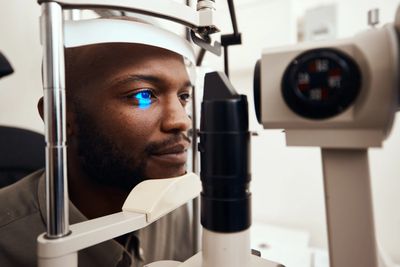
Below are some of the eye conditions our doctors diagnose and manage as part of our comprehensive eye care services, emphasizing the early detection of eye diseases to ensure optimal vision preservation.
Retina and Macula Conditions
Plaquenil (Hydroxychloroquine) Retinal Toxicity
Hydroxychloroquine (Plaquenil) is an effective medication used in comprehensive eye care to treat autoimmune diseases such as lupus and rheumatoid arthritis. Although retinal toxicity is uncommon, prolonged use or higher cumulative doses may increase the risk of permanent retinal damage. Regular screening with specialized testing, including Optical Coherence Tomography (OCT) and visual field testing, is crucial for the early detection of eye diseases, allowing for the recognition of retinal changes before noticeable vision loss occurs.
→ Learn More About Plaquenil (Hydroxychloroquine) Retinal Toxicity
Age-Related Macular Degeneration (AMD)
Age-related macular degeneration impacts the macula, the central portion of the retina responsible for sharp, detailed vision, complicating activities like reading, driving, and recognizing faces. Early detection and regular monitoring are vital for vision preservation.
→ Learn More About Age-Related Macular Degeneration (AMD)
Diabetic Retinopathy
Diabetic retinopathy occurs when diabetes damages the small blood vessels of the retina, making it one of the leading causes of vision loss among adults with diabetes. Routine diabetic eye examinations are essential for detecting retinal changes before symptoms develop, aiding in early detection and proactive management.
→ Learn More About Diabetic Retinopathy
Diabetic Macular Edema (DME)
Diabetic macular edema, a complication of diabetic retinopathy, leads to fluid accumulation within the macula, resulting in blurred or distorted central vision. Early diagnosis and management are critical for vision preservation.
→ Learn More About Diabetic Macular Edema (DME)
Posterior Vitreous Detachment (PVD)
A posterior vitreous detachment occurs when the vitreous gel naturally separates from the retina as part of the aging process. While common, a PVD can occasionally lead to retinal tears, making prompt evaluation of any new flashes or floaters essential in comprehensive eye care.
→ Learn More About Posterior Vitreous Detachment (PVD)
Retinal Tears
A retinal tear develops when the retina is pulled or torn, often during a posterior vitreous detachment. Without timely evaluation and treatment when indicated, a retinal tear may progress to a retinal detachment, underscoring the need for comprehensive monitoring.
→ Learn More About Retinal Tears
Retinal Detachment
A retinal detachment occurs when the retina separates from the back of the eye. Symptoms may include flashes of light, numerous new floaters, or a curtain-like shadow over the vision. This condition requires prompt medical evaluation to ensure vision preservation.
→ Learn More About Retinal Detachment
Floaters
Floaters are common visual symptoms that may be normal or may indicate a retinal condition requiring urgent evaluation. Sudden increases in these symptoms should always be examined promptly, reflecting the importance of comprehensive eye care.
Epiretinal Membrane (Macular Pucker)
An epiretinal membrane is a thin layer of scar-like tissue that forms on the surface of the macula, potentially leading to blurred or distorted central vision. Regular monitoring is important for early detection and management options.
→ Learn More About Epiretinal Membrane (Macular Pucker)
Macular Hole
A macular hole is a small opening that develops in the center of the macula, often resulting in blurred or distorted central vision, particularly when reading or performing detailed tasks. Early intervention can aid in the preservation of vision.
→ Learn More About Macular Hole
Vitreomacular Traction (VMT)
Vitreomacular traction occurs when the vitreous remains partially attached to the macula, placing traction on retinal tissue. This may lead to blurred vision or distortion, highlighting the need for comprehensive eye care and monitoring to prevent further complications.
→ Learn More About Vitreomacular Traction (VMT)
Central Serous Chorioretinopathy (CSCR)
Central serous chorioretinopathy occurs when fluid accumulates beneath the retina, often causing blurred or distorted central vision. Many cases improve over time, but some may require additional evaluation or treatment, emphasizing the importance of early detection of eye diseases.
→ Learn More About Central Serous Chorioretinopathy (CSCR)
Retinal Vein Occlusion (RVO)
A retinal vein occlusion occurs when one of the veins draining blood from the retina becomes blocked. Depending on the severity, patients may experience sudden or gradual vision loss, necessitating prompt ophthalmic evaluation for optimal outcomes.
→ Learn More About Retinal Vein Occlusion (RVO)
Retinal Artery Occlusion (RAO)
A retinal artery occlusion occurs when blood flow through a retinal artery is interrupted, often causing sudden, painless vision loss. This is a medical emergency requiring immediate evaluation to ensure the greatest chance of vision preservation.
→ Learn More About Retinal Artery Occlusion (RAO)
Cystoid Macular Edema (CME)
Cystoid macular edema occurs when fluid collects within the layers of the macula. This condition may develop after eye surgery or due to inflammation or other retinal diseases, leading to blurred central vision. Regular monitoring and timely management are essential in comprehensive eye care.
→ Learn More About Cystoid Macular Edema (CME)
Lattice Degeneration
Lattice degeneration refers to a thinning of the peripheral retina, increasing the risk of retinal tears in some individuals. While most patients have no symptoms, routine retinal examinations play a key role in monitoring changes and promoting early detection of eye diseases.
→ Learn More About Lattice Degeneration
Hypertensive Retinopathy
Hypertensive retinopathy occurs when high blood pressure damages the retinal blood vessels. Comprehensive eye examinations can reveal retinal changes that reflect overall vascular health, highlighting the importance of blood pressure control in vision preservation.
→ Learn More About Hypertensive Retinopathy
Retinitis Pigmentosa
Retinitis pigmentosa encompasses a group of inherited retinal disorders that gradually affect night vision and peripheral vision. Regular monitoring is essential to assess disease progression, maximize remaining vision, and discuss available treatment options and supportive care.
→ Learn More About Retinitis Pigmentosa
Ocular Toxoplasmosis
Ocular toxoplasmosis is caused by Toxoplasma gondii, a microscopic parasite that infects millions of people worldwide.
Most individuals who carry the parasite never know they have it. In many cases, the infection occurs years earlier and remains dormant within the body without causing symptoms.
→ Learn More About Ocular Toxoplasmosis
Optic Nerve Conditions
Glaucoma
Glaucoma is a group of eye diseases that damage the optic nerve and stands as one of the leading causes of irreversible vision loss worldwide. Because glaucoma often develops without noticeable symptoms, comprehensive eye care, including routine examinations, is essential for the early detection of eye diseases and long-term monitoring.
Optic Neuritis
Optic neuritis is the inflammation of the optic nerve that can lead to sudden vision loss, decreased color vision, and eye pain, particularly during eye movement. This condition may arise independently or be linked with neurological conditions such as multiple sclerosis, necessitating prompt evaluation.
→ Learn More About Optic Neuritis
Ischemic Optic Neuropathy
Ischemic optic neuropathy occurs when blood flow to the optic nerve is reduced, leading to sudden vision loss. Depending on the underlying cause, this condition may be related to vascular disease or inflammatory disorders and requires prompt medical assessment for effective vision preservation.
→ Learn More About Ischemic Optic Neuropathy
Optic Atrophy
Optic atrophy refers to the degeneration of the optic nerve as a result of previous injury or disease. Depending on the underlying cause, patients may experience reduced vision, decreased color perception, or visual field loss, highlighting the importance of ongoing comprehensive eye care.
→ Learn More About Optic Atrophy
Papilledema
Papilledema is the swelling of the optic nerve caused by increased pressure within the skull. It may come with symptoms such as headaches, vision changes, nausea, or other neurological signs, making urgent medical evaluation necessary to determine the underlying cause.
→ Learn More About Papilledema
Optic Disc Drusen
Optic disc drusen are calcified deposits within the optic nerve head that could resemble optic nerve swelling during an eye examination. While many individuals may have no symptoms, optic disc drusen can occasionally impact peripheral vision and should be monitored regularly as part of comprehensive eye care.
→ Learn More About Optic Disc Drusen
Congenital Optic Nerve Anomalies
Some individuals are born with structural differences of the optic nerve, such as optic nerve hypoplasia or optic nerve coloboma. These congenital conditions vary in severity and may affect vision, making comprehensive eye examinations crucial for diagnosis and long-term monitoring to support vision preservation.
→ Learn More About Congenital Optic Nerve Anomalies
Corneal Conditions
Keratoconus
Keratoconus is a progressive eye condition where the cornea thins and gradually bulges into a cone-like shape. This irregular curvature can lead to blurred or distorted vision, glare, and heightened sensitivity to light. Early detection of eye diseases is crucial, as treatment options differ depending on the disease's stage. Comprehensive eye care is essential for managing this condition effectively.
→ Learn More About Keratoconus
Corneal Abrasion
A corneal abrasion is a scratch on the clear front surface of the eye. Symptoms typically include pain, tearing, redness, sensitivity to light, blurred vision, and the feeling of something in the eye. Prompt evaluation is vital to reduce the risk of infection and support proper healing, thereby aiding in vision preservation.
→ Learn More About Corneal Abrasion
Corneal Ulcer
A corneal ulcer presents as an open sore on the cornea, often resulting from infection, contact lens complications, or eye injury. Symptoms may involve severe eye pain, redness, blurred vision, discharge, and light sensitivity. Corneal ulcers require prompt medical attention to help prevent permanent vision loss, emphasizing the importance of early detection of eye diseases.
→ Learn More About Corneal Ulcer
Recurrent Corneal Erosion
Recurrent corneal erosion occurs when the outer layer of the cornea repeatedly separates from the underlying tissue, typically following an eye injury or due to specific corneal disorders. Episodes can cause sudden eye pain, tearing, blurred vision, and light sensitivity, especially upon waking. Comprehensive eye care can help manage these recurrences effectively.
→ Learn More About Recurrent Corneal Erosion
Fuchs’ Endothelial Corneal Dystrophy
Fuchs’ dystrophy is an inherited condition affecting the innermost layer of the cornea. As the disease progresses, fluid builds up within the cornea, resulting in blurred vision, glare, and increased sensitivity to light. Early detection of eye diseases and consistent monitoring are critical for vision preservation.
→ Learn More About Fuchs’ Endothelial Corneal Dystrophy
Corneal Dystrophies
Corneal dystrophies encompass a group of inherited disorders that impact one or more layers of the cornea. Depending on the specific type, they may lead to blurred vision, recurrent corneal erosions, or discomfort. Many progress slowly and benefit from regular monitoring and comprehensive eye care.
→ Learn More About Corneal Dystrophies
Corneal Edema
Corneal edema refers to swelling of the cornea caused by excess fluid within its layers. It may arise following eye surgery, an injury, elevated eye pressure, or certain corneal diseases, resulting in blurred vision, glare, or halos around lights. Early detection enables effective management and enhances vision preservation.
→ Learn More About Corneal Edema
Corneal Scars
Corneal scarring can develop after infections, injuries, ulcers, or previous eye surgeries. Depending on the size and location of the scar, it may disrupt the passage of light through the cornea and influence visual clarity, underscoring the importance of comprehensive eye care.
→ Learn More About Corneal Scars
Corneal Degenerations
Corneal degenerations are generally age-related or environmentally influenced changes impacting the cornea. Unlike corneal dystrophies, these changes are typically not inherited and may gradually develop over time. Some cases may require only observation, while others could affect vision or comfort, highlighting the need for regular assessments as part of comprehensive eye care.
→ Learn More About Corneal Degenerations
Infectious Keratitis
Infectious keratitis is an infection of the cornea caused by bacteria, viruses, fungi, or parasites. This condition is considered an ocular emergency, as delayed diagnosis or treatment can lead to permanent vision loss. Contact lens wear is a common risk factor for certain types of infectious keratitis, making early detection of eye diseases critical.
→ Learn More About Infectious Keratitis
Eyelid & Ocular Surface Conditions
Dry Eye Disease
Dry eye disease occurs when your eyes do not produce enough healthy tears or when tears evaporate too quickly. Symptoms may include burning, stinging, fluctuating vision, redness, excessive tearing, and a gritty sensation. This condition is common and can significantly impact comfort and quality of life, making comprehensive eye care essential for managing symptoms and ensuring overall eye health.
→ Learn More About Dry Eye Disease
Meibomian Gland Dysfunction (MGD)
Meibomian gland dysfunction is one of the most prevalent causes of dry eye disease. It occurs when the oil-producing glands along the eyelid margins become blocked or do not function properly, causing tears to evaporate too quickly and leading to irritation, redness, and fluctuating vision. Early detection of eye diseases like MGD can help improve treatment outcomes and comfort.
→ Learn More About Meibomian Gland Dysfunction (MGD)
Blepharitis
Blepharitis is inflammation of the eyelid margins that may cause redness, burning, itching, crusting around the eyelashes, and irritation. It is often a chronic condition that can contribute to dry eye symptoms and requires ongoing eyelid care to preserve vision and prevent further complications.
→ Learn More About Blepharitis
Demodex Blepharitis
Demodex blepharitis is a form of blepharitis caused by an overgrowth of microscopic Demodex mites that naturally live around the eyelashes. Symptoms may include itching, irritation, redness, crusting, and the presence of cylindrical dandruff around the lashes. Regular comprehensive eye care can help identify and manage this condition effectively.
→ Learn More About Demodex Blepharitis
Conjunctivitis (Pink Eye)
Conjunctivitis, commonly known as pink eye, is inflammation of the conjunctiva, the thin membrane covering the white part of the eye and the inner eyelids. It may be caused by viral or bacterial infections, allergies, or irritation. Treatment depends on identifying the underlying cause, highlighting the importance of early detection of eye diseases.
→ Learn More About Conjunctivitis (Pink Eye)
Eye Allergies (Allergic Conjunctivitis)
Eye allergies occur when the eyes react to allergens such as pollen, pet dander, dust mites, or mold. Common symptoms include itching, redness, tearing, and swelling. Identifying and managing the underlying trigger can help improve comfort and ensure effective vision preservation.
→ Learn More About Eye Allergies
Pinguecula
A pinguecula is a common, non-cancerous yellowish growth on the conjunctiva, usually located near the cornea. It is often associated with long-term exposure to sunlight, wind, or dry environments and may occasionally become inflamed or irritated, necessitating a comprehensive eye care approach.
→ Learn More About Pinguecula
Pterygium
A pterygium is a wedge-shaped growth of tissue that extends from the conjunctiva onto the cornea. It is commonly linked to ultraviolet (UV) light exposure and may cause irritation, redness, or blurred vision if it grows over the visual axis. Early detection of eye diseases like pterygium is vital for treatment and management.
→ Learn More About Pterygium
Stye (Hordeolum)
A stye is an acute infection of an eyelid gland that typically appears as a painful, swollen bump near the edge of the eyelid. Most styes improve with conservative treatment, but persistent or recurrent cases should be evaluated by an eye care professional for proper management and vision preservation.
→ Learn More About Styes (Hordeola)
Chalazion
A chalazion is a painless lump that develops when one of the eyelid’s oil glands becomes blocked. Unlike a stye, a chalazion is usually not caused by an active infection, although it may develop after one. Comprehensive eye care can address chalazia effectively.
→ Learn More About Chalazia
Giant Papillary Conjunctivitis (GPC)
Giant papillary conjunctivitis is an inflammatory condition most commonly associated with contact lens wear, ocular prostheses, or exposed sutures. Symptoms may include itching, excessive lens movement, mucus discharge, and reduced contact lens comfort. Early detection of this condition can improve patient comfort significantly.
→ Learn More About Giant Papillary Conjunctivitis (GPC)
Conjunctival Cysts
Conjunctival cysts are fluid-filled sacs that develop on the conjunctiva. Most are benign and painless, although larger cysts may cause irritation, a foreign body sensation, or cosmetic concerns. Regular eye examinations are essential for monitoring these cysts as part of diligent comprehensive eye care.
→ Learn More About Conjunctival Cysts
Lens Conditions
Cataracts
A cataract occurs when the eye’s natural lens becomes cloudy, leading to blurred vision, glare, reduced contrast sensitivity, and difficulty seeing at night. While cataracts typically develop gradually due to aging, they can also result from eye injuries, certain medications, or other medical conditions. To ensure early detection of eye diseases such as cataracts, regular comprehensive eye care examinations are essential in determining when treatment may be beneficial.
→ Learn More About Cataracts
Congenital Cataracts
Congenital cataracts are present at birth or develop during early childhood. Depending on their size and location, these cataracts can interfere with normal visual development, emphasizing the need for prompt evaluation to preserve vision and reduce the risk of permanent vision problems.
→ Learn More About Congenital Cataracts
Traumatic Cataracts
Traumatic cataracts develop following an injury to the eye. Vision changes may manifest immediately after the injury or gradually over time, depending on the severity and type of trauma. Comprehensive eye care after such incidents is crucial to monitor and address these changes effectively.
→ Learn More About Traumatic Cataracts
Posterior Capsule Opacification (Secondary Cataract)
Posterior capsule opacification (PCO), often referred to as a “secondary cataract,” is a common condition that can develop months or years after cataract surgery. It results in blurry vision similar to a cataract, but it is not a true cataract. Fortunately, vision can often be restored through a quick, painless laser procedure performed by an ophthalmologist, ensuring ongoing vision preservation.
→ Learn More About Posterior Capsule Opacification
Ectopia Lentis (Dislocated Lens)
Ectopia lentis is a condition where the eye’s natural lens is displaced from its normal position. This can happen due to trauma, inherited disorders, or connective tissue diseases, significantly affecting vision based on the degree of lens displacement. Regular comprehensive eye care can help monitor this condition.
→ Learn More About Ectopia Lentis
Presbyopia
Presbyopia is a normal age-related change where the eye gradually loses its ability to focus on nearby objects. Most people notice difficulty reading small print or using digital devices in their early to mid-40s. Glasses, contact lenses, or other vision correction options can enhance near vision, contributing to overall vision preservation.
→ Learn More About Presbyopia
Inflammatory & Autoimmune Eye Conditions Uveitis
Uveitis
Uveitis is inflammation inside the eye that can affect the iris, ciliary body, or choroid. Symptoms may include eye pain, redness, blurred vision, light sensitivity, and floaters. For comprehensive eye care, prompt diagnosis and treatment are crucial to reduce the risk of permanent vision loss.
→ Learn More About Uveitis
Iritis (Anterior Uveitis)
Iritis is the most common form of uveitis, involving inflammation of the iris, the colored part of the eye. Symptoms often develop suddenly and may include eye pain, redness, blurred vision, and sensitivity to light. Early evaluation and treatment are essential for the early detection of eye diseases, helping to prevent complications.
→ Learn More About Iritis
Episcleritis
Episcleritis is inflammation of the thin layer of tissue between the conjunctiva and the white part of the eye. It typically causes redness and mild discomfort but is generally less painful and less serious than scleritis. Although many cases resolve on their own, an eye examination is recommended for early detection and to confirm the diagnosis.
→ Learn More About Episcleritis
Scleritis
Scleritis is a serious inflammatory condition affecting the white outer wall of the eye (sclera). It often causes severe eye pain, redness, light sensitivity, and decreased vision. Scleritis may be associated with autoimmune diseases, making prompt medical evaluation and treatment vital for vision preservation.
→ Learn More About Scleritis
Ocular Rosacea
Ocular rosacea is a chronic inflammatory condition that affects the eyelids and surface of the eyes. Symptoms may include burning, dryness, redness, tearing, and recurrent styes or blepharitis. Many people with ocular rosacea also experience skin rosacea on their face, necessitating regular eye assessments for early detection and management of potential complications.
→ Learn More About Ocular Rosacea
Thyroid Eye Disease
Thyroid eye disease is an autoimmune disorder commonly associated with Graves’ disease. It can cause eye bulging, eyelid retraction, dryness, double vision, eye discomfort, and, in severe cases, vision loss. Early diagnosis and appropriate management are crucial for protecting eye health and aiding in vision preservation.
→ Learn More About Thyroid Eye Disease
Ocular Manifestations of Autoimmune Disease
Autoimmune diseases such as rheumatoid arthritis, lupus, Sjögren’s syndrome, ankylosing spondylitis, inflammatory bowel disease, and psoriasis can affect the eyes in various ways, including causing dry eye, uveitis, scleritis, and other inflammatory conditions. Regular eye examinations are essential for the early detection of eye diseases and effective management.
→ Learn More About Autoimmune Eye Disease
Sjögren’s Syndrome and Dry Eye
Sjögren’s syndrome is an autoimmune disease that commonly affects the tear and salivary glands, often resulting in significant dry eye disease and dry mouth. Early recognition and appropriate management can improve comfort and protect the surface of the eyes, making comprehensive eye care vital for those affected.
→ Learn More About Sjögren’s Syndrome & Dry Eye
Eye Injuries and Emergencies
Foreign Body in the Eye
Dust, metal, wood, glass, or other particles can become lodged in or on the surface of the eye, causing pain, tearing, redness, light sensitivity, and the sensation that something is in the eye. Prompt evaluation is essential for safe removal of the foreign body, minimizing the risk of infection and preserving vision. This is an important aspect of comprehensive eye care.
→ Learn More About Foreign Bodies in the Eye
Corneal Abrasion
A corneal abrasion is a scratch on the clear front surface of the eye. It commonly results in significant pain, tearing, redness, blurred vision, and light sensitivity. Early detection and treatment are vital to prevent infection and support proper healing, facilitating the early detection of eye diseases.
→ Learn More About Corneal Abrasion
Chemical Eye Injuries
Chemical injuries occur when acids, alkalis, or other irritating substances come into contact with the eye. Immediate irrigation followed by prompt medical evaluation is critical. Early treatment can considerably improve the chances of preserving vision and is vital in any instance of chemical exposure.
→ Learn More About Chemical Eye Injuries
Blunt Eye Trauma
Blunt trauma to the eye can stem from sports injuries, falls, motor vehicle accidents, or other impacts. Depending on severity, it may lead to bruising, bleeding inside the eye, retinal injuries, fractures around the eye, or vision loss. A comprehensive eye examination is necessary after any significant eye injury to ensure early detection of potential issues.
→ Learn More About Blunt Eye Trauma
Penetrating Eye Injuries
Penetrating or perforating eye injuries happen when an object enters the eye. These injuries are true ocular emergencies that require immediate evaluation to reduce the risk of permanent vision loss and other serious complications, underscoring the importance of vision preservation in eye health.
→ Learn More About Penetrating Eye Injuries
Hyphema
Hyphema is bleeding into the front chamber of the eye, usually following blunt trauma. Symptoms may include pain, blurred vision, and visible blood inside the eye. Prompt evaluation and careful monitoring are necessary, as complications can affect vision and eye pressure, highlighting the need for comprehensive eye care.
→ Learn More About Hyphema
Orbital Fractures
An orbital fracture involves a break in one or more of the bones surrounding the eye, often due to facial trauma. Symptoms may include swelling, bruising, double vision, numbness, or difficulty moving the eye. Evaluation is critical to determine whether additional treatment or surgical consultation is needed, reinforcing the importance of early detection of eye diseases.
→ Learn More About Orbital Fractures
Flashes & Floaters
A sudden increase in flashes of light or floaters may indicate a retinal tear or detachment, particularly if accompanied by a curtain-like shadow or loss of peripheral vision. These symptoms should always be evaluated promptly to preserve vision.
→ Learn More About Flashes & Floaters
Sudden Vision Loss
Sudden vision loss may arise from various causes including retinal disease, optic nerve disorders, vascular conditions, or other medical emergencies. Any sudden decrease in vision should be treated as an emergency and assessed as soon as possible for effective vision preservation.
→ Learn More About Sudden Vision Loss
Red Eye
While many red eye cases stem from minor conditions such as allergies or irritation, redness can also indicate serious eye diseases or infections. Accompanying symptoms like eye pain, decreased vision, or light sensitivity warrant prompt evaluation.
→ Learn More About Red Eye
Eye Pain
Eye pain can range from mild irritation to severe discomfort caused by infection, inflammation, injury, elevated eye pressure, or other serious conditions. Persistent or severe eye pain should not be disregarded and deserves prompt evaluation within the framework of comprehensive eye care.
→ Learn More About Eye Pain
Sudden Double Vision
Sudden double vision (diplopia) may suggest issues with eye muscles, nerves, brain, or blood vessels. Given that some causes can be quite serious, sudden double vision should be assessed quickly to ensure timely intervention.
→ Learn More About Sudden Double Vision
Vision & Neurological Conditions
Ocular Migraine
An ocular migraine can cause temporary visual disturbances such as flashing lights, zigzag patterns, shimmering lights, blind spots, or blurred vision. While these symptoms usually resolve on their own, they should be evaluated by a professional, especially if they are new, prolonged, or accompanied by other neurological symptoms. Comprehensive eye care is crucial for understanding these occurrences.
→ Learn More About Ocular Migraine
Visual Aura (Migraine Aura)
A visual aura is a temporary neurological symptom that often precedes or accompanies a migraine. Patients may notice flashing lights, geometric patterns, blind spots, or distorted vision. Because similar symptoms may occur with retinal or other neurological conditions, new visual auras should be evaluated by an eye care professional to ensure the early detection of eye diseases.
→ Learn More About Visual Aura
Double Vision (Diplopia)
Double vision occurs when a person sees two images of a single object. It may stem from issues involving the eye muscles, nerves, cornea, lens, or brain. Due to the potential seriousness of some causes, persistent or sudden double vision should be evaluated promptly for effective vision preservation.
→ Learn More About Double Vision (Diplopia)
Nystagmus
Nystagmus is an involuntary, repetitive movement of the eyes that can be present from birth or develop later in life. Depending on the underlying cause, it can affect vision, depth perception, and balance, all of which are crucial for effective vision preservation.
→ Learn More About Nystagmus
Cranial Nerve Palsies
The nerves controlling eye movement can become weakened or damaged due to diabetes, vascular disease, trauma, or neurological conditions. Cranial nerve palsies may lead to double vision, eyelid drooping, or difficulty moving the eyes and often require prompt evaluation and comprehensive eye care.
→ Learn More About Cranial Nerve Palsies
Visual Field Defects
Visual field defects are areas of reduced or missing vision, potentially resulting from glaucoma, retinal disease, optic nerve disorders, stroke, brain tumors, or other neurological conditions. Specialized testing in the context of comprehensive eye care can help identify and monitor these defects for the early detection of eye diseases.
→ Learn More About Visual Field Defects
Homonymous Hemianopia
Homonymous hemianopia is the loss of one-half of the visual field in both eyes, typically caused by a stroke, brain injury, or another neurological condition affecting the visual pathways. This condition can significantly impact reading, driving, and daily activities, underscoring the importance of vision preservation.
→ Learn More About Homonymous Hemianopia
Cortical Visual Impairment
Cortical visual impairment occurs when vision loss results from damage to the brain's visual processing centers instead of the eyes themselves. Symptoms vary depending on the location and severity of the neurological injury, highlighting the need for comprehensive eye care.
→ Learn More About Cortical Visual Impairment
Transient Vision Loss (Amaurosis Fugax)
Transient vision loss is a temporary loss of vision in one or both eyes that may last from seconds to minutes. Given that it can be a warning sign of vascular or neurological disease, prompt medical evaluation is essential for both the early detection of eye diseases and overall vision preservation.
→ Learn More About Transient Vision Loss
Visual Snow Syndrome
Visual snow syndrome is a neurological condition where individuals continuously see tiny flickering dots throughout their visual field, often accompanied by other visual symptoms. A comprehensive eye examination helps rule out underlying eye disease before a neurological diagnosis is considered, emphasizing the importance of early detection of eye diseases.
→ Learn More About Visual Snow Syndrome
Eye Muscle & Binocular Vision Conditions
Strabismus (Eye Turn)
Strabismus is a condition in which the eyes do not align properly and point in different directions. This misalignment can be present from childhood or develop later in life due to neurological conditions, trauma, or other eye disorders. Early detection of eye diseases, such as strabismus, is crucial as it can significantly improve visual function and eye alignment.
→ Learn More About Strabismus
Amblyopia (Lazy Eye)
Amblyopia, commonly referred to as “lazy eye,” happens when one eye fails to develop normal vision during childhood. The early detection and treatment of this condition are vital since vision preservation is generally most successful while the visual system is still developing.
→ Learn More About Amblyopia (Lazy Eye)
Convergence Insufficiency
Convergence insufficiency is a binocular vision disorder that makes it difficult for the eyes to work together when focusing on nearby objects. Common symptoms include eye strain, headaches, blurred vision, double vision, and trouble reading or concentrating during near tasks. Comprehensive eye care can help identify and manage this condition effectively.
→ Learn More About Convergence Insufficiency
Divergence Insufficiency
Divergence insufficiency is a binocular vision disorder causing challenges for the eyes to coordinate when looking at distant objects. Patients frequently experience double vision while driving or viewing faraway objects. Timely intervention is important for optimal outcomes.
→ Learn More About Divergence Insufficiency
Convergence Excess
Convergence excess occurs when the eyes turn inward more than needed during near tasks. This may lead to headaches, eye strain, blurred vision, or intermittent double vision while reading or using digital devices. Comprehensive eye care can help manage this issue effectively.
→ Learn More About Convergence Excess
Accommodative Dysfunction
Accommodative dysfunction pertains to difficulties in focusing clearly on objects at various distances. This condition may cause blurred vision, headaches, eye fatigue, or trouble maintaining clear vision during extended reading or computer use. Early detection of eye diseases will aid in treatment options for those affected.
→ Learn More About Accommodative Dysfunction
Diplopia (Double Vision)
Diplopia occurs when a person sees two images of a single object. This condition may arise from problems involving the eye muscles, nerves controlling eye movement, or other neurological or ocular issues. Sudden or persistent double vision should always be evaluated promptly to prevent further complications.
→ Learn More About Diplopia (Double Vision)
Nystagmus
Nystagmus is characterized by involuntary, repetitive eye movements that can reduce visual acuity and affect depth perception. It can be congenital or acquired later in life and may be associated with neurological or ocular conditions. Comprehensive eye care is vital for those experiencing this disorder.
→ Learn More About Nystagmus
Cranial Nerve Palsies
Damage to the cranial nerves responsible for controlling eye movement can lead to double vision, abnormal eye alignment, eyelid drooping, or difficulty in moving the eyes. These issues often require prompt evaluation to determine the underlying cause and initiate appropriate care.
→ Learn More About Cranial Nerve Palsies
Fourth Nerve Palsy
A fourth cranial nerve palsy impacts the superior oblique muscle, commonly causing vertical double vision, especially when reading or walking down stairs. This disorder may be congenital or acquired due to trauma, vascular disease, or other neurological conditions. Early detection of eye diseases in this context can enhance treatment success.
→ Learn More About Fourth Nerve Palsy
Sixth Nerve Palsy
A sixth cranial nerve palsy weakens the lateral rectus muscle, making it challenging to move the eye outward. Patients often report horizontal double vision that worsens when looking towards the affected side. Comprehensive eye care is essential for managing this condition.
→ Learn More About Sixth Nerve Palsy
Internuclear Ophthalmoplegia (INO)
Internuclear ophthalmoplegia is a neurological eye movement disorder resulting from damage to the pathways connecting the eye movement centers within the brain. It typically leads to impaired horizontal eye movements and double vision. Early detection and treatment are crucial for vision preservation in affected individuals.
→ Learn More About Internuclear Ophthalmoplegia (INO)
Refractive Conditions
Myopia (Nearsightedness)
Myopia, or nearsightedness, is a common refractive condition where distant objects appear blurry while nearby objects remain clear. This condition occurs when light focuses in front of the retina rather than directly on it. Myopia can often be corrected with glasses, contact lenses, or other vision correction options. Routine comprehensive eye care is essential for identifying and managing myopia effectively.
→ Learn More About Myopia (Nearsightedness)
Hyperopia (Farsightedness)
Hyperopia, or farsightedness, occurs when light focuses behind the retina, making nearby objects difficult to see clearly. Depending on the degree of hyperopia and a person’s age, both near and distance vision may be affected. Treatment options typically include glasses or contact lenses. Early detection of eye diseases related to hyperopia can significantly enhance vision preservation.
→ Learn More About Hyperopia (Farsightedness)
Astigmatism
Astigmatism is a common refractive condition caused by an irregular curvature of the cornea or lens. Instead of focusing light evenly on the retina, the eye produces blurred or distorted vision at all distances. Glasses, contact lenses, and various other vision correction options can effectively improve vision. Comprehensive eye care can aid in diagnosing astigmatism early to support vision preservation.
→ Learn More About Astigmatism
Presbyopia
Presbyopia is a normal age-related change in focusing ability that typically begins in the early to mid-40s. As the eye's natural lens becomes less flexible, reading small print and focusing on nearby objects become more challenging. Reading glasses, multifocal lenses, and other vision correction options can help restore clear near vision. Regular eye examinations facilitate the early detection of eye diseases associated with presbyopia.
Anisometropia
Anisometropia occurs when the two eyes have significantly different refractive errors. Depending on the degree of difference, it may cause blurred vision, difficulty with depth perception, eye strain, or amblyopia (lazy eye), particularly in children. Comprehensive eye care is vital for recognizing and managing anisometropia effectively.
→ Learn More About Anisometropia
Aniseikonia
Aniseikonia is a condition in which each eye perceives images at different sizes. It may result from significant differences in prescription, certain eye conditions, or previous eye surgery, leading to eyestrain, headaches, double vision, or difficulty with depth perception. Early detection through comprehensive eye care can aid in vision preservation.
→ Learn More About Aniseikonia
Progressive Myopia
Progressive myopia refers to nearsightedness that continues to worsen over time, especially during childhood and adolescence. Increasing levels of myopia may heighten the risk of certain eye diseases later in life, making regular eye examinations and appropriate myopia management crucial for long-term vision preservation.
→ Learn More About Progressive Myopia
Pathologic Myopia
Pathologic myopia is a severe form of nearsightedness characterized by excessive elongation of the eye, which increases the risk of retinal disease, glaucoma, cataracts, and other vision-threatening complications. Regular comprehensive eye examinations are essential for monitoring long-term eye health and facilitating the early detection of eye diseases associated with pathologic myopia.
→ Learn More About Pathologic Myopia